The rise of antibiotic-resistant bacteria has initiated a quest for
alternatives to conventional antibiotics. One potential alternative is
PlyC, a potent enzyme that kills the bacteria that causes strep throat
and streptococcal toxic shock syndrome. PlyC operates by locking onto
the surface of a bacteria cell and chewing a hole in the cell wall large
enough for the bacteria’s inner membrane to protrude from the cell,
ultimately causing the cell to burst and die.
Research has shown
that alternative antimicrobials such as PlyC can effectively kill
bacteria. However, fundamental questions remain about how bacteria
respond to the holes that these therapeutics make in their cell wall and
what size holes bacteria can withstand before breaking apart. Answering
those questions could improve the effectiveness of current
antibacterial drugs and initiate the development of new ones.
Researchers
at the Georgia Institute of Technology and the University of Maryland
recently conducted a study to try to answer those questions. The
researchers created a biophysical model of the response of a
Gram-positive bacterium to the formation of a hole in its cell wall.
Then they used experimental measurements to validate the theory, which
predicted that a hole in the bacteria cell wall larger than 15 to 24
nanometers in diameter would cause the cell to lyse, or burst. These
small holes are approximately one-hundredth the diameter of a typical
bacterial cell.
“Our model correctly predicted that the membrane
and cell contents of Gram-positive bacteria cells explode out of holes
in cell walls that exceed a few dozen nanometers. This critical hole
size, validated by experiments, is much larger than the holes
Gram-positive bacteria use to transport molecules necessary for their
survival, which have been estimated to be less than 7 nanometers in
diameter,” said
Joshua Weitz,
an associate professor in the School of Biology at Georgia Tech. Weitz
also holds an adjunct appointment in the School of Physics at Georgia
Tech.
The study was published online on Jan. 9, 2013 in the
Journal of the Royal Society Interface. The work was supported by the James S. McDonnell Foundation and the Burroughs Wellcome Fund.
Common Gram-positive bacteria that infect humans include
Streptococcus, which causes strep throat;
Staphylococcus, which causes impetigo; and
Clostridium, which causes botulism and tetanus. Gram-negative bacteria include
Escherichia, which causes urinary tract infections;
Vibrio, which causes cholera; and
Neisseria, which causes gonorrhea.
Gram-positive
bacteria differ from Gram-negative bacteria in the structure of their
cell walls. The cell wall constitutes the outer layer of Gram-positive
bacteria, whereas the cell wall lies between the inner and outer
membrane of Gram-negative bacteria and is therefore protected from
direct exposure to the environment.
Georgia Tech biology graduate
student Gabriel Mitchell, Georgia Tech physics professor Kurt Wiesenfeld
and Weitz developed a biophysical theory of the response of a
Gram-positive bacterium to the formation of a hole in its cell wall. The
model detailed the effect of pressure, bending and stretching forces on
the changing configuration of the cell membrane due to a hole. The
force associated with bending and stretching pulls the membrane inward,
while the pressure from the inside of the cell pushes the membrane
outward through the hole.
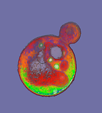
 |
A transmission electron microscope image of a Streptococcus
pyogenes cell experiencing lysis after exposure to the
highly active enzyme PlyC. (Credit: Daniel Nelson, UMD) |
“We found that bending forces act to
keep the membrane together and push it back inside, but a sufficiently
large hole enables the bending forces to be overpowered by the internal
pressure forces and the membrane begins to escape out and the cell
contents follow,” said Weitz.
The balance between the bending and
pressure forces led to the model prediction that holes 15 to 24
nanometers in diameter or larger would cause a bacteria cell to burst.
To test the theory,
Daniel Nelson,
an assistant professor at the University of Maryland, used transmission
electron microscopy images to measure the size of holes created in
lysed
Streptococcus pyogenes bacteria cells following PlyC exposure.
Nelson
found holes in the lysed bacteria cells that ranged in diameter from 22
to 180 nanometers, with a mean diameter of 68 nanometers. These
experimental measurements agreed with the researchers’ theoretical
prediction of critical hole sizes that cause bacterial cell death.
According to the researchers, their theoretical model is the first to consider the effects of cell wall thickness on lysis.
“Because
lysis events occur most often at thinner points in the cell wall, cell
wall thickness may play a role in suppressing lysis by serving as a
buffer against the formation of large holes,” said Mitchell.
The
combination of theory and experiments used in this study provided
insights into the effect of defects on a cell’s viability and the
mechanisms used by enzymes to disrupt homeostasis and cause bacteria
cell death. To further understand the mechanisms behind enzyme-induced
lysis, the researchers plan to measure membrane dynamics as a function
of hole geometry in the future.
source:http://www.gatech.edu/newsroom/release.html?nid=182231