TEST

Sunday, 11 November 2012

First ‘snapshots’ of the electronic structure of a manganese complex related to water-splitting in photosynthesis

Together with a large international research team, Johannes Messinger of Umeå University in Sweden has taken another step toward an understanding of photosynthesis and developing artificial photosynthesis. With a combination of a x-ray free-electron laser and spectroscopy, the team has managed to see the electronic structure of a manganese complex, a chemical compound related to how photosynthesis splits water.
 illustration of ultra-short x-ray pulse striking molecules
containing manganese. Illustration: Greg Stewart,
National Accelerator Laboratory at Stanford University
The experiments used the Linac Coherent Light Source (LCLS), which is a free-electron x-ray laser facility at Stanford University in the US. The wavelength of the laser is roughly the same as the breadth of an atom, and each pulse of light lasts 50 femtoseconds (10-15). This is an extremely short interval of time: there are more femtoseconds in one second than there are seconds in a person’s life. Such extremely short wavelengths and short light pulses constitute ideal conditions for imaging chemical reactions with atomic resolution at room temperature while the chemical reactions are ongoing.
The research group has previously used LCLS to perform structural analyses of isolated photosynthesis complexes from plants’ photosystem II at room temperature. Now the group has combined the method with spectroscopy and is the first team to succeed in seeing at LCLS the electronic structure of a manganese complex similar to that found in photosystem II. Manganese is a transitional metal that, together with calcium and oxygen, forms the water-splitting catalyst in photosystem II.
A very simple example of a spectrometer is a prism, which separates sunlight into all the colors of the rainbow. The spectrometer used in this study functions in a similar manner, but with a group of 16 specialized crystals that diffract the x-rays emitted from the sample in resonse of being excited by an x-ray pulse onto a detector array.
To the delight of the scientists, the manganese compounds remained intact long enough for them to observe detailed information about the electronic structure before the compounds were destroyed by the very intense X-ray laser beam.
“Having both structural information and spectroscopic information means that we can much better understand how the structural changes of the whole complex and the chemical changes on the active surface of the catalysts work together to enable the enzymes to perform complex chemical reactions at room temperature,” says Johannes Messinger, professor at the Department of Chemistry at Umeå University.
The chemical reaction the research group aims to understand is the splitting of water in photosystem II, as this understanding is also key for developing artificial photosynthesis– that is, for building devices for producing hydrogen from sunlight and water. To be able to exploit sunlight for producing fuels that can be stored and the used when needed would help solve the world’s ever-more acute energy problems.
source:http://www.teknat.umu.se/english/about-the-faculty/news/newsdetailpage/first-snapshots-of-the-electronic-structure-of-a-manganese-complex-related-to-water-splitting-in-photosynthesis.cid202064

read more

Wyss Institute Models a Human Disease in an Organ-on-a-Chip

Researchers at the Wyss Institute for Biologically Inspired Engineering at Harvard University have mimicked pulmonary edema in a microchip lined by living human cells, as reported today in the journal Science Translation Medicine. They used this "lung-on-a-chip" to study drug toxicity and identify potential new therapies to prevent this life-threatening condition.

 The study offers further proof-of-concept that human "organs-on-chips" hold tremendous potential to replace traditional approaches to drug discovery and development.
"Major pharmaceutical companies spend a lot of time and a huge amount of money on cell cultures and animal testing to develop new drugs," says Donald Ingber, M.D., Ph.D., founding director of the Wyss Institute and senior author of the study, "but these methods often fail to predict the effects of these agents when they reach humans."
The lung-on-a-chip device [see video], which the team first described only two years ago, is a crystal clear, flexible polymer about the size of a memory stick that contains hollow channels fabricated using computer microchip manufacturing techniques. Two of the channels are separated by a thin, flexible, porous membrane that on one side is lined with human lung cells from the air sac and exposed to air; human capillary blood cells are placed on the other side with medium flowing over their surface. A vacuum applied to side channels deforms this tissue-tissue interface to re-create the way human lung tissues physically expand and retract when breathing.

Wyss Technology Development Fellow Dongeun Huh, Ph.D., who also holds appointments at Boston Children's Hospital and Harvard Medical School, studied a cancer chemotherapy drug called interleukin-2 -- or IL-2 for short -- in the lung-on-a-chip. A major toxic side effect of IL-2 is pulmonary edema, which is a deadly condition in which the lungs fill with fluid and blood clots.


 When IL-2 was injected into the blood channel of the lung-on-a-chip, fluid leaked across the membrane and two tissue layers, reducing the volume of air in the other channel and compromising oxygen transport -- just as it does in lungs of human patients when it is administered at the equivalent doses and over the same time course. Blood plasma proteins also crossed into the air channel, leading to the formation of blood clots in the air space, as they do in humans treated with IL-2.
But one result came as a surprise.
It turns out the physical act of breathing greatly enhances the effects of IL-2 in pulmonary edema -- "something that clinicians and scientists never suspected before," Ingber says. When the team turned on the vacuum attached to the chip to simulate breathing, it increased fluid leakage more than three-fold when treated with the clinically relevant IL-2 dose, and the Wyss team confirmed that the same response occurs in an animal model of pulmonary edema. This result could suggest that doctors treating patients on a respirator with IL-2 should reduce the tidal volume of air being pushed into the lungs, for example, in order to minimize the negative side effects of this drug.
Combining microfabrication techniques with modern
tissue engineering, lung-on-a-chip offers an in
vitro approach to drug screening by mimicking
the complicated mechanical and biochemical
behaviors of a human lung.
Most exciting for the future of drug testing was the Wyss team's finding that "this on-chip model of human pulmonary edema can be used to identify new potential therapeutic agents in vitro," Ingber says. The pulmonary edema symptoms in the lung-on-a-chip disease model could be prevented by treating the tissues with a new class of drug, a transient receptor potential vanilloid 4 (TRPV4) channel blocker, under development by GlaxoSmithKline (GSK). In a separate study published by the GSK team in the same issue of Science Translation Medicine, the beneficial effects of TRPV4 inhibition in reducing pulmonary edema were independently validated using animal models of pulmonary edema caused by heart failure.
"In just a little more than two years, we've gone from unveiling the initial design of the lung-on-a-chip to demonstrating its potential to model a complex human disease, which we believe provides a glimpse of what drug discovery and development might look like in the future," Ingber says.

source:http://wyss.harvard.edu/viewpressrelease/99/


read more

Monday, 5 November 2012

Biochemists Discover New Mechanism in Ribosome Formation

A new mechanism in the formation of ribosomes has been discovered by researchers from the Heidelberg University Biochemistry Center. In an interdisciplinary approach, the Heidelberg scientists, along with colleagues from Switzerland and Japan, describe a heretofore uncharacterised protein that plays a specific role in ribosome assembly in eukaryotes, organisms whose cells contain a cell nucleus. This protein makes sure that specific factors required for ribosome synthesis are transported together, like hitchhikers, into the nucleus to the site of assembly. The results of this research were published in “Science”.
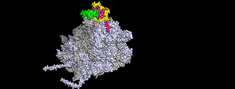
The figure shows the large subunit of the ribosome in its high-resolution 3D
structure. The ribosomal RNA is depicted in grey, the myriad of ribosomal proteins
in blue-grey. The r-protein Rpl5 is shown in yellow, the r-protein Rpl11 in green. The
ruby-coloured area reveals the ribosomal RNA which binds Rpl5 and Rpl11.

Ribosomes, the protein factories of the cell, are macromolecular complexes of ribonucleic acids (RNA) and ribosomal proteins (r-proteins) that are organised in a highly complicated three-dimensional nanostructure. Correct synthesis of ribosomes is critical for the division of all cells and is a process that follows strict rules. In eukaryotes, new ribosomes are formed predominantly in the cell nucleus. Therefore, the r-proteins needed for ribosome formation must travel from the cytoplasm of the cell to a site in the nucleus where the ribosomes are assembled. Until recently it was not clear whether r-proteins that have a similar function and form functional clusters on the ribosome structure are also co-transported into the nucleus.
The researchers have now found a protein that coordinates the co-transport of certain r-proteins in functional clusters into the cell nucleus. This factor is called Symportin1, for synchronised import. “Symportin1 synchronises the import of both the Rpl5 and Rpl11 r-proteins into the cell nucleus and supports their integration into the growing ribosome structure”, explains Prof. Dr. Irmgard Sinning of the Heidelberg University Biochemistry Center (BZH). “It employs a familiar logistical concept from every day life, like picking up a hitchhiker or sharing a taxi with someone headed for the same destination”, says Dr. Gert Bange of the BZH, lead author of the study together with Dr. Dieter Kressler (now of Fribourg University).
source: http://www.uni-heidelberg.de/presse/news2012/pm201211102_ribosomen_en.html
read more

NYU researchers use simulations on TACC, XSEDE supercomputers to understand how some carcinogens evade removal by stabilizing the very DNA they attack

A person doesn't have to go far to find a polycyclic aromatic hydrocarbon (PAH). These carcinogen precursors are inhaled through automobiles exhaust during the morning commute, are present in a drag of cigarette smoke, and are part of any barbequed meal.

Once ingested or inhaled, these big, bulky multi-ringed molecules are converted into reactive carcinogenic compounds that can bind to DNA, sometimes literally bending the double helix out of its normal shape, to form areas of damage called lesions. The damaged DNA can create errors in the genetic code during replication, which may cause cancer-initiating mutations.
It is the job of the nuclear excision repair (NER) system to repair damage caused by PAH lesions by removing the segment of DNA where the lesion is bound and patching up the resulting gap. But some lesions are especially resistant to this repair machinery, making them much more likely to cause mutations than lesions that are promptly repaired.
Models showing the steric hinderance between different lesions (colored yellow and turquoise)and DNA. Steric hinderance causes double helix destabilization and refers to the degree of crowding between different atoms.
A research team at New York University (NYU) has gained new insight on the ability of certain PAH-derived lesions to evade the DNA repair machinery. They found that some lesions stabilize the DNA they damage, making it difficult for a certain repair protein to mark the lesion for repair. Their research was published earlier this year in the February 2012 issue of Biochemistry. More recent articles about NER of DNA lesions from the same group appeared in Nucleic Acids Research in July and August.
"Some lesions cause DNA to be locally destabilized, but there are lesions that actually stabilize the DNA so that the two strands come apart with great difficulty," said Suse Broyde, a biology professor at NYU. "Sometimes they're even more stable than undamaged DNA."
The stability of the DNA double helix is a key feature that determines whether DNA is flagged for repair in the first place by a protein called XPC. The protein patrols the genome looking for weakened areas. When it finds one, it slips a structure called a beta-hairpin between the strands, marking the DNA for NER. But if a lesion makes DNA more stable, the strands become more difficult to separate and the beta hairpin can't signal for repair.
How can molecules as large and disruptive as a PAH stabilize DNA in the first place?


"If you remember from chemistry, every kind of molecule interacts with other molecules through so-called Van der Waals interactions," said Nicholas Geacintov, a chemistry professor at NYU. "The DNA and the carcinogen bound to it also have the same kind of interactions," he said, specifically referring to the PAH-derived lesions that intercalate, or wedge themselves, between DNA base pairs.
The role of Van der Waals forces, which Broyde calls "stacking interactions" for these systems, was made clear through a series of computer simulations performed, analyzed, interpreted and visualized by Yuqin Cai, a post-doctoral senior research scientist in Broyde's lab. "The computer simulations revealed the structural, energetic and dynamic properties of the DNA containing the PAH-derived lesions, " said Broyde, who specializes in providing a mechanistic understanding of complex biological processes using molecular dynamics simulations and other front-line computational approaches.
"You can make movies of the dynamic trajectory which allow you to see the real mobility of the entire system," Broyde said. "You can watch the DNA flexing and the backbone moving dynamically and the carcinogen moving in and out. It's not rigid — you can see its aliveness."

Nuclear Excision Repair of a PAH-derived lesion.
Animation courtesy of Yuqin Cai.

Broyde and her team's simulations revealed that of the six different lesions examined (three chemicals with two different geometric configurations each), those caused by dibenzo[a,l]pyrene, the most tumorigenic PAH investigated, were the most resistant to repair. The five-ringed structure of the carcinogen provided ample stacking opportunities, which stabilized the DNA much better than the four and three-ringed structures of the other PAHs that were examined.

Knowing which lesions are the most repair resistant could play an important role in preventative medicine, said Broyde, as individuals harboring them could be counseled to avoid further exposure, particularly in the case of smokers.
The atomic-level visualizations Broyde's lab simulated explained data gathered from experiments that are carried out in Geacintov's lab, including synthesis of the damaged DNA, measurements of its stability, and investigations of the relative NER susceptibility of lesion-containing DNA in human cells.
"Without experiments they wouldn't have anything to model and without modeling it would be very hard for us to understand what we are measuring," Geacintov said.
Some of the most important results came from DNA melting experiments, where a specific sequence of double-stranded DNA was synthesized with a known lesion and then heated up until the strands separated. The more stable the DNA, the higher the temperature at which it melted. The most stabilizing lesion melted nearly 10 degrees higher than the melting temperature of DNA with no lesions; this lesion was the most repair-resistant in experiments and also most stabilizing according to molecular simulations.

Suse Broyde is a biology professor at New York University.

To compute the raw data (coordinates of structures as a function of time) for the analyses and visualizations, the Broyde lab used the Longhorn, Lonestar and Ranger high-performance computing resources at the Texas Advanced Computing Center as well as other systems in the Extreme Science and Engineering Discovery Environment (XSEDE).
Uncovering the stabilizing properties that allow some lesions to evade NER and initiate cancer could help develop better chemotherapeutic drugs to fight the disease, according to Broyde and Geacintov.
For example, the widely used chemotherapy drug, Cisplatin, attacks the DNA of cancerous cells, interrupting unregulated replication. However, the cell's own NER machinery can combat the drug by removing it and repairing the genome, just as in healthy non-cancerous cells.
"One direction in drug design is to find pharmaceuticals that still inhibit replication but that are less susceptible to NER," Broyde said. "Understanding the mechanism of NER will be valuable in designing the next generation of chemotherapeutic agents. These would be more effective if they were more resistant to NER."

source :Texas Advanced Computing Center
read more

Friday, 2 November 2012

Ames Lab researchers find three unique cell-to-cell bonds

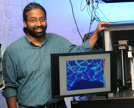
Sanjeevi Sivasankar leads a research team that uses atomic force
microscopy and other technologies to study the bonds that connect
biological cells.  Photo by Bob Elbert.
The human body has more than a trillion cells, most of them connected, cell to neighboring cells.
How, exactly, do those bonds work? What happens when a pulling force is applied to those bonds? How long before they break? Does a better understanding of all those bonds and their responses to force have implications for fighting disease?
Sanjeevi Sivasankar, an Iowa State assistant professor of physics and astronomy and an associate of the U.S. Department of Energy’s Ames Laboratory, is leading a research team that’s answering those questions as it studies the biomechanics and biophysics of the proteins that bond cells together.
The researchers discovered three types of bonds when they subjected common adhesion proteins (called cadherins) to a pulling force: ideal, catch and slip bonds. The three bonds react differently to that force: ideal bonds aren’t affected, catch bonds last longer and slip bonds don’t last as long.
The findings have just been published by the online Early Edition of the Proceedings of the National Academy of Sciences.
Sivasankar said ideal bonds – the ones that aren’t affected by the pulling force – had not been seen in any previous experiments. The researchers discovered them as they observed catch bonds transitioning to slip bonds.
“Ideal bonds are like a nanoscale shock absorber,” Sivasankar said. “They dampen all the force.”
And the others?
“Catch bonds are like a nanoscale seatbelt,” he said. “They become stronger when pulled. Slip bonds are more conventional; they weaken and break when tugged.”
In addition to Sivasankar, the researchers publishing the discovery are Sabyasachi Rakshit, an Iowa State post-doctoral research associate in physics and astronomy and an Ames Laboratory associate; Kristine Manibog and Omer Shafraz, Iowa State doctoral students in physics and astronomy and Ames Laboratory student associates; and Yunxiang Zhang, a post-doctoral research associate for the University of California, Berkeley’s California Institute for Quantitative Biosciences.
The project was supported by a $308,000 grant from the American Heart Association, a $150,000 Basil O’Connor Award from the March of Dimes Foundation and Sivasankar’s Iowa State startup funds.
The researchers made their discovery by taking single-molecule force measurements with an atomic force microscope. They coated the microscope tip and surface with cadherins, lowered the tip to the surface so bonds could form, pulled the tip back, held it and measured how long the bonds lasted under a range of constant pulling force.
The researchers propose that cell binding “is a dynamic process; cadherins tailor their adhesion in response to changes in the mechanical properties of their surrounding environment,” according to the paper.
When you cut your finger, for example, cells filling the wound might use catch bonds that resist the pulls and forces placed on the wound. As the forces go away with healing, the cells may transition to ideal bonds and then to slip bonds.
Sivasankar said problems with cell adhesion can lead to diseases, including cancers and cardiovascular problems.
And so Sivasankar said the research team is pursuing other studies of cell-to-cell bonds: “This is the beginning of a lot to be discovered about the role of these types of interactions in healthy physiology as well as diseases like cancer.”
source:http://www.news.iastate.edu/news/2012/11/01/cellbonds
read more

New Technique Enables High-Sensitivity View of Cellular Functions

Tiny amounts of carbohydrates (1 zmol, correspnding to a few hundred molecules) can be detected quantitatively by a real-time method based on the conjugation of carbohydrates with DNA marker. The  method called glyco-qPCR uses amplification to provide uniform, ultrasensitive detection of carbohydrates, which can be applied to glycobiology, as well as carbohydrate-based drug discovery.
Researchers at Rensselaer Polytechnic Institute have developed an ultrasensitive method for detecting sugar molecules — or glycans — coming from living organisms, a breakthrough that will make possible a more detailed understanding of cellular functions than either genetic or proteomic (the study of proteins) information can provide. The researchers hope the new technique will revolutionize the study of glycans, which has been hampered by an inability to easily detect and identify minute quantities of these molecules.
“The glycome is richer in information than the genome or the proteome. A cancer cell, for example, might have the same genome as a non-cancer cell, but it produces different sugars,” said Robert Linhardt, the Ann and John H. Broadbent Jr. ’59 Senior Constellation Professor of Biocatalysis and Metabolic Engineering at Rensselaer, and an author of the study. “Until now, the stumbling block in glycomics has been rapid and sensitive determination of the glycans present in a biological sample, and up to now we were very limited by how much we could detect. With this technique that we’ve developed, Glyco-qPCR, we can detect a very small number of molecules and that should accelerate the growth of the field.”
The new technique is discussed in a paper titled “Signal Amplification by Glyco-qPCR for Ultrasensitive Detection of Carbohydrates: Applications in Glycobiology,” which was published in the Oct. 16 online edition of Angewandte Chemie International. Linhardt and Jonathan Dordick, director of the Rensselaer Center for Biotechnology and Interdisciplinary Studies (CBIS), vice president for research, and the Howard P. Isermann ’42 Professor of Chemical and Biological Engineering, were joined in the research by Seok Joon Kwon, Kyung Bok Lee, Kemal Solakyildirim, Sayaka Masuko, Mellisa Ly, Fuming Zhang, and Lingyn Li.
Linhardt used the analogy of a house to explain the importance of glycans in biology and the promise of glycomics in medicine and biotechnology: If genes are the blueprints, and proteins are the structure, than sugars—glycans—are the decoration of all living matter. Just as dozens of houses in a development—despite a shared blueprint and identical external appearance—can have a unique interior identity based on wall colors and furnishings, so can two cells share the same genome, and similar proteome, but function very differently from one another, Linhardt explained.
“You can look at a blueprint of a house and it can tell you something about the house, but it certainly can’t tell you the colors of the walls,” Linhardt said. “We’ve developed a method to start to detect what the decorations will look like, and that will give us an insight into what the house will ultimately become.”
Linhardt said the technique is likely to find applications in the study of all complex multisystem diseases, such as cancer and diabetes.
“This gives us a new tool to study fundamental biology and chemistry,” Linhardt said. “It allows us a higher resolution view into the functions of a cell than the genome or proteome. With this tool we can go inside a cell, poke around, and understand how to predict the behavior of that cell and ultimately control it.”
As the name of the new technique suggests, Glyco-qPCR is built on Polymerase Chain Reaction (PCR), a technique, which enabled fast and cost-effective sequencing of genetic information, fueling a rapid expansion in genetics starting in the mid-1980s.
PCR allows researchers to produce mass copies of a particular sequence of DNA, or “amplify” the sequence, turning one precious sample into a nearly limitless supply of a particular sequence. The large sample makes it possible to perform other techniques that determine the identity of the particular sequence.
Glycans, the sugar molecules present in living cells, are even smaller and more complex than DNA sequences, and therefore, even more difficult to identify, Linhardt said. Moreover, unlike DNA, they have proven resistant to “amplification.” So the Linhardt team took another approach.
The team has developed a technique for chemically attaching a specific DNA sequence to a specific sugar molecule. The team has built a catalogue of molecules that can be “tagged,” each with a specific DNA sequence.
Once tagged, the team uses PCR to amplify the DNA tags, allowing them to identify the tags — and therefore the glycans — that are present, and the proportions in which they are present, in a given sample.
“We don’t really detect the molecule, we detect the DNA that’s attached to it,” Linhardt said. “The DNA tags are cleverly designed so that they only attach to certain molecules. We can then amplify the DNA, see what kind of DNA it is, and then infer the molecule that it’s attached to.”
None of the currently used methods of glycan analysis, such as mass spectrometry or high-performance liquid chromatography, amplify the amount of sample that is present so they are much less sensitive, Linhardt said. While these current methods are capable of detecting a few billion glycan molecules, Glyco-qPCR can detect a few hundred glycan molecules.
The development of PCR in 1983 put the study of genes within reach of research labs around the world, unlocking the potential for knowledge about how genes work and treatments build on that knowledge. Linhardt hopes Glyco-qPCR will effect a similar transformation.
 “Although it is an indirect method that piggy-backs on PCR, amplification technology like our Glyco-qPCR holds the same promise for glycomics research,” Linhardt said. “I believe that it is revolutionary for the fields of glycomics and glycobiology.”

source: http://news.rpi.edu/update.do?artcenterkey=3103
read more

Friday, 26 October 2012

Academia Should Fulfill Social Contract by Supporting Bioscience Startups, Case Study Says

Universities not only provide the ideal petri dish for cultivating bioscience with commercial potential, but have a moral obligation to do so, given the opportunity to translate public funding into health and jobs, according to a new case study by UCSF researchers.

In an analysis published Wednesday in Science Translational Medicine, researchers at the California Institute for Quantitative Biosciences (QB3) assessed the impact of the institute’s efforts over the past eight years in supporting entrepreneurs on the three UC campuses in which it operates: UCSF, UC Berkeley and UC Santa Cruz.

The study found that, by lowering the hurdle even very slightly for scientists to become entrepreneurs, the scientists were able to gain extraordinary traction in translating academic research into public benefit, generating 60 new companies in the first six years and attracting 75 new bioscience entrepreneurs in the last year alone.
“This fundamentally changes the way we think of academic science,” said Douglas Crawford, PhD, assistant director of QB3 and senior author on the paper. “There is a distressing paucity of new drugs in the pipeline and a clear need for new economic engines in this country. This is a call to action to address that.”
The paper cites the following essential support that universities can provide:
  • Bioscience-focused incubators
  • An open network approach that enables any entrepreneur to participate
  • Competitive seed funding options
  • Real-world mentoring that gives scientist a clear sense of market needs.
At UCSF, this support is happening on multiple fronts, starting with the efforts underway at QB3, which focus on supporting entrepreneurs directly. UCSF also is home to the National Institute of Health's first Clinical and Translational Sciences Institute (CTSI), which has become a model nationwide in creating a vast network of scientists in both clinical and basic research, in an effort to improve collaboration between them and expedite translational research to improve health. CTSI also is developing broad coalitions and partnerships at the local and national levels to enable a transformation of the research environment. In addition, UCSFs' office of Innovation, Technology and Alliances is a leader nationwide in supporting entrepreneurship on campus, while forging direct collaborations with industry.

Growing Incubators of Bioscience Companies
In the first six years since QB3 started supporting entrepreneurs at the UCSF Mission Bay campus, its growing network of incubators helped launch 60 new bioscience companies at UCSF and across the San Francisco Bay at UC Berkeley — at a cost of $1 million per year. Of those, 56 are still in business, and 13 have moved beyond the incubator network or have been purchased by larger companies.
Together, those companies have created more than 280 jobs and attracted more than $230 million in either small business grants or venture capital funding for those companies, which are primarily focused on developing therapeutics, medical devices and research tools. That’s a 38-fold return on investment, despite current negative trends in seed-stage investing, in addition to the public benefit of any products those companies generate.


The federal government invests $50 billion per year in academic research, including $23 billion for life sciences from the National Institutes of Health (NIH) alone, according to the paper. Both Baltimore, which is home to universities, and the San Francisco Bay Area receive about $2 billion annually in government research and development funding, yet productivity as measured by high-tech startups was 20-fold higher in the San Francisco area than Baltimore, the analysis found.
(A) Regional differences in R&D spending. R&D spending by universities (left axis)
and number of companies listed in the Deloitte Technology Fast 500 (right axis) in
selected urban centers. Analysis by the Center for an Urban Future, prepared with
data from the National Science Foundation/Division of Science Resources
Statistics. (B) Growth of the QB3 Garage Network. Millions of dollars invested in
QB3 Garage Network companies each year (left axis), and number of new
start-ups that joined the QB3 Garage Network per year (right axis). (C) Number
and types of jobs created in the QB3 Garage Network.
CREDIT: C. BICKEL/SCIENCE TRANSLATIONAL MEDICINE

“This is clearly not an issue of how much funding is coming to the region. It’s an issue of how we think about our social contract,” said Regis B. Kelly, PhD, UCSF professor of biochemistry and biophysics, executive director of QB3 and a co-author on the paper. “Most people would say the public good a university offers is in providing research and education, but if we truly want to support the public with the benefits of science, we need to stop seeing economic growth and new medications as an inadvertent byproduct of academic research.”
Many U.S. universities, including UCSF, are exploring ways to work with the private sector, with the multiple goals of invigorating research on campus, expediting drug development and ultimately improving the therapies that make it to the market.
That’s a relatively new way of thinking, though, and one that often has been met with both skepticism and charges of conflicts of interest by those who believe science should be completely devoid of commercial influences.


“We have a fundamental charge as scientists to make an economic impact and to translate science into public benefit,” Crawford said. “That’s not a traditional way of thinking of universities, but at a certain point, we need to reassess that traditional thinking and take pride in the fact that a university’s public contributions extends beyond its classrooms.”
The paper points to the mission statement of the NIH, the nation’s largest bioscience funding agency, which specifically includes a goal “to enhance the Nation’s economic well-being and ensure a continued high return on the public investment in research.”
“So as academic scientists, we are actually paid to protect and improve health, to prevent disease, and to enhance economic well-being,” Crawford said. “This is not a sell-out to industry. It’s an obligation to society.”

Crawford emphasized that the goal of the analysis is not to assess blame, but to find solutions, and there are many. The authors also hope to generate discussion about which solutions work.


source : http://www.ucsf.edu/news/2012/10/13005/academia-should-fulfill-social-contract-supporting-bioscience-startups-case-study
read more

Whitehead scientists identify major flaw in standard approach to global gene expression analysis

The assumption in traditional genetic expression analysis that mRNA content is similar between cells (represented by orange and black dots) does not affect the final results when the cells in fact do have equivalent mRNA content, as in Figure A.  In Figure B, the cell represented by the orange dots has a significantly higher mRNA content, but when the data is normalized with the assumption that their contents are equal, the perceived response is skewed and inaccurately indicates that some of the genes are repressed (green bars).  Using a standardized control, as in Figure C, eliminates the assumptions about mRNA content and presents accurate results.
Whitehead Institute researchers report that common assumptions employed in the generation and interpretation of data from global gene expression analyses can lead to seriously flawed conclusions about gene activity and cell behavior in a wide range of current biological research.
“Expression analysis is one of the most commonly used methods in modern biology,” says Whitehead Member Richard Young. “So we are concerned that flawed assumptions may affect the interpretation of many biological studies.”
Much of today’s interpretation of gene expression data relies on the assumption that all cells being analyzed have similar total amounts of messenger RNA (mRNA), the roughly 10% of a cell’s RNA that acts as a blueprint for protein synthesis. However, some cells, including aggressive cancer cells, produce several times more mRNA than other cells. Traditional global gene expression analyses have typically ignored such differences.
“We’ve highlighted this common assumption in gene expression analysis that potentially affects many researchers,” says Tony Lee, a scientist in Young’s lab and a corresponding author of the article published in this week’s issue of Cell. “We provided a concrete example of the problem and a solution that can be implemented by investigators.”
Members of the Young lab recently uncovered the flaw while investigating genes expressed in cancer cells expressing high levels of c-Myc, a gene regulator known to be highly expressed in aggressive cancer cells.  When comparing cells with high and low c-Myc levels, they were surprised to find very different results using different approaches to gene expression analysis.  Further investigation revealed that there were striking differences in the total amounts of RNA from the high and low c-Myc -containing cells, yet these differences were masked by commonly used experimental and analytical methods.
“The different results we saw from different methods of gene expression analysis were shocking, and led us to reinvestigate the whole process on several platforms,” says Jakob Lovén, postdoctoral reseacher in Young’s lab and co-author of the Cell paper. “We then realized that the common assumption that cells contain similar levels of mRNA is badly flawed and can lead to serious misinterpretations, particularly with cancer cells that can have very different amounts of RNA.”
In addition to delineating this problem, the Whitehead scientists also describe a remedy. By using synthetically produced mRNAs, called RNA spike-ins, as standardized controls, researchers can compare experimental data and eliminate assumptions about total cell RNA amounts.  The remedy applies to all three gene expression analysis platforms they studied.
Although the researchers believe the use of RNA spike-ins should become the new standard for global gene expression analyses, questions are likely to persist about the interpretations of much prior research.
“There are over 750,000 expression datasets in public databases, and because they generally lack information about the cell numbers used in the analysis, it is unclear whether they can be re-examined in order to validate the original interpretation” says David Orlando, a scientist in the Young lab. “It may be necessary to reinvestigate some important concepts.”

source: http://wi.mit.edu/news/archive/2012/whitehead-scientists-identify-major-flaw-standard-approach-global-gene-expression
read more

Tuesday, 23 October 2012

A new glow for electron microscopy

In this electron microscope image of a mitochondrion, the matrix has
been stained with APEX, making it appear dark. The lighter
projections into the matrix represent the intermembrane space.
Image: Tom Deerinck and Jeff Martell
The glowing green molecule known as green fluorescent protein (GFP) has revolutionized molecular biology. When GFP is attached to a particular protein inside a cell, scientists can easily identify and locate it using fluorescence microscopy. However, GFP can’t be used with electron microscopy, which offers much higher resolution than fluorescence microscopy.

Chemists from MIT have now designed a GFP equivalent for electron microscopy — a tag that allows scientists to label and visualize proteins with unprecedented clarity.

“With things that may appear only a few pixels across by fluorescence microscopy — for example, a mitochondrion — you can’t make out any of the internal features. But with electron microscopy it’s very easy to discern the intricate internal structures,” says Jeff Martell, a graduate student in chemistry at MIT and lead author of a paper describing the new tag in the Oct. 21 online edition of Nature Biotechnology.

The new tag could help scientists pinpoint the locations of many cell proteins, providing new insight into those proteins’ functions, according to the researchers.

Improving on nature

Dubbed APEX, the new tag is similar to naturally occurring proteins that have been tried as imaging labels for electron microscopy. Horseradish peroxidase (HRP) is one commonly used tag, but it works only in a few compartments of a cell. Other recently developed tags work throughout a cell but are technically challenging to use because they require light to be shined on the sample and oxygen to be bubbled through it.

To improve on these methods, the researchers started with a protein similar to HRP, called ascorbate peroxidase (APX). APX is more versatile than HRP because it can function within a cell’s cytosol, in the main cavity of a cell.

Both HRP and APX belong to a class of enzymes called peroxidases, which remove an electron and a proton from other molecules in a process known as oxidation. Every peroxidase has different targets, and one of HRP’s main targets is a molecule called DAB, which when oxidized can be visualized with electron microscopy. The researchers genetically engineered APX so that it would also target DAB.

To use this new APEX tag (for “engineered APX”), the researchers deliver, into a living cell, a small ring of DNA containing the APEX gene joined to the gene for the protein they plan to image. The cell then produces the target protein, bound to the APEX protein.

Next, the researchers need to deliver DAB, which is not normally found in cells. This delivery takes place during the process of “fixing,” or stabilizing cells, which must be done before they can be imaged with electron microscopy.

When the APEX protein oxidizes DAB, it generates radicals that rapidly clump together into a tarlike polymer. That polymer can be detected through electron microscopy, allowing the researchers to pinpoint the location of the target protein.

A biological question resolved

To demonstrate the usefulness of their new tag, the researchers set out to resolve an open question regarding the location of a calcium channel protein discovered last year. Two research groups identified the protein and reported that it is located within mitochondria, but they had conflicting theories as to its precise location and orientation. Using the new imaging technique, the MIT-led team labeled the protein and determined that it is embedded in the inner mitochondrial membrane and faces into the innermost part of mitochondria, the mitochondrial matrix.

The team also showed that the new tag can label proteins throughout the cell — not only within mitochondria but also in the nucleus, the endoplasmic reticulum and the cytosol.

In current studies, the researchers are working on filling entire cells, such as neurons, with their imaging agent. This allows certain neurons in an electron microscope image to stand out, making it easier to trace the connections they make with other neurons.

“We want to find the exact connections that these cells are making, and APEX is a good way to label cells for electron microscopy. We can label specific types of cells and figure out how they fit into the neural circuitry,” Sanes says.


source : http://web.mit.edu/newsoffice/2012/a-new-glow-for-electron-microscopy-1021.html
read more