-->

TEST

Saturday, 27 April 2013

Application for Junior Research Fellow (JRF) and AcSIR Ph.D. Programme August 2013 at CSIR-IICT, Hyderabad

CSIR-INDIAN INSTITUTE OF CHEMICAL TECHNOLOGY
(Council of Scientific & Industrial Research)
Hyderabad 500 607
Application for Junior Research Fellow (JRF) and AcSIR Ph.D. Programme
CSIR-IICT-PhD – August 2013

CSIR-IICT invites applications from highly talented and research oriented candidates to join as Junior Research Fellow (JRF) to pursue AcSIR PhD Program at CSIR-IICT in the following areas, for the Academic Session starting from August 2013.

Research Area :

Natural Products
Crop Protection Chemicals
Organic & Bimolecular Chemistry
Fluoroorganics
Lipids Science and Technology
Medicinal Chemistry & Pharmacology
Analytical Chemistry
Molecular Modelling
Polymers & Functional Materials
Inorganic & Physical Chemistry
Biology/ Chemical Biology
Bioengineering and Environmental Sciences
Chemical Engineering

Eligibility : Candidates must fulfil any one of the following eligibility criteria:
1. First class M.Sc. degree or equivalent in Chemistry/ Biology/ Allied sciences With
i) a valid rank in CSIR/UGC-NET JRF Examination, OR
ii) a valid DST INSPIRE/ICMR/ Fellowship.
Note : The candidates who have passed CSIR-UGC-NET examination, under the Lectureship category only are NOT eligible
2. First class BE/B.Tech in Chemical Engineering with a valid GATE score / CSIR-UGC-NET rank
Note : The candidates who have passed CSIR-UGC-NET examination, under the Lectureship category only are NOT eligible

AcSIR PhD Registration : All JRFs selected for the above Program shall be registered under the Academy of Scientific and Innovative Research (AcSIR) for pursing their PhD. Hence the candidates must necessarily submit on-line application to AcSIR at http://acsir.res.in/on or before May 23, 2013

Fellowship : Candidates selected for the PhD Program at CSIR-IICT shall draw their fellowship and other allowances, contingency etc., as per their respective fellowship schemes.

Application procedure :
1. Candidates who fulfil the eligibility criteria may apply online for PhD Admission to CSIR- IICT at www.iictindia.org on or before May 23, 2013
2. In addition, the candidates MUST ALSO submit on-line applications for PhD registration in AcSIR at http://acsir.res.in/ on or before May 23, 2013

PLEASE MAIL A SOFT COPY OF THE RECEIPT/ACKNOWLEDGEMENT OF AcSIR to phdatiict@gmail.com

Selection : Selection for admission to the Ph.D. Programme shall be on the basis of interview(s)/ Written Test to be conducted at CSIR-IICT for the short-listed candidates. The interviews are likely to be conducted on June 17 – 18, 2013. Please note that as per the applicable norms no TA/DA shall be paid to the candidates appearing for the interview/ Written Test.

Deadline : 23.05.13

read more

ISCA YOUNG SCIENTIST'S PROGRAM 2013

To encourage Young Scientists, The Indian Science Congress Association has instituted a number of awards in different disciplines. These awards carry a sum of Rs.25,000/- besides a Certificate of Merit. Applications are invited from members (Life &Annual) of the Association who have paid their subscription on or before July 15, 2013. The upper age limit of the candidates for the award is 32 years as reckoned on December 31, 2013.

Four copies of the abstract (not exceeding 100 words) along with four copies of full length paper must reach the office of the General Secretary (Membership Affairs) not later than August 16, 2013. At the top of each copy of the paper and its abstract, the name of the Section under which the paper is to be considered should be indicated. For details of Sections seehttp://www.sciencecongress.nic.in/html/youngsc.htmlare 

Along with the Four copies of paper, Four copies of the Application Form (to be downloaded from ISCA websitehttp://www.sciencecongress.nic.in/ ) with brief bio-data of the candidate (not exceeding 2 pages), list of publications , with copies of reprints of already published papers if any and a soft copy of the duly filled application form with scanned copies of enclosures (excluding reprints), full length paper, abstract and bio data in the form of a CD must also be sent simultaneously along with the hard copies. 

The Paper submitted must be a single author paper and the research work should have been carried out in India and this has to be certified by the Head of the Institution from where the candidate is applying. The candidate should give an undertaking that the paper being submitted has not been published in any journal or presented in any other Conference / Seminar / Symposium or submitted for consideration of any award. A Young Scientist can present only one paper in any one Section (and not a second paper on the same or any other topic in any other Section).
A person who has already received Young Scientist Award in any section once will not be eligible to apply for the above Award in the same or any other section. Incomplete Applications will not be considered. The papers submitted will be subjected to verification for authenticity . Full length paper will be evaluated by experts and the selected Six Young Scientists in each section will be invited to make oral presentation of their paper during 101st Indian Science Congress. 

The selected candidates will be provided admissible travelling allowances by the ISCA. The final selection for the Awards will be made by a duly constituted committee and the awards will be given during the Valedictory Session of 101st Indian Science Congress session. Applications submitted for the above award will not be returned. The last date for receiving papers at ISCA Headquarters is August 16, 2013.

All Correspondences should be made to: 
The General Secretary (Membership Affairs.), 
The Indian Science Congress Association, 14,
Dr. Biresh Guha St., Kolkata-700017. 
Tel. Nos. (033) 2287-4530/2281-5323
Fax No.91-33-2287-2551 E-mail : iscacal@vsnl.net 
Website : http://www.sciencecongress.nic.in/ Download Application Form

read more

Thursday, 25 April 2013

HAPPY DNA DAY

Today is DNA Day, a holiday commemorating the day in 1953 when James Watson, Francis Crick, Maurice Wilkins, Rosalind Franklin and various colleagues all published papers on the structure of DNA.

This discovery revolutionized biology and was without a doubt one of the most important scientific discoveries of the last 100 years.

All of their papers are now freely available for the public to read. Celebrate DNA day by reading the original research articles, and by passing them along for friends to read.

Watson, James Dewey; Crick, Francis Harry Compton (1953-04-25). "Molecular structure of nucleic acids; a structure for deoxyribose nucleic acid". Nature 171 (4356): 737–738. Read here: http://bit.ly/13asBk

Franklin, Rosalind Elsie; Gosling, Raymond (1953-04-25). "Molecular configuration in sodium thymonucleate". Nature 171 (4356): 740–741. Read it here: http://bit.ly/hf4eaz

Wilkins, Maurice Hugh Frederick; Stokes, Alexander Rawson; Wilson, Herbert R. (1953-04-25). "Molecular structure of deoxypentose nucleic acids". Nature 171 (4356): 738–740. Read it here: http://bit.ly/17WK6dI

read more

Thursday, 14 March 2013

Can Fungi Replace Plastics? Maybe, Say Researchers


Fungi, with the exception of shitake and certain other mushrooms, tend to be something we associate with moldy bread or dank-smelling mildew. But they really deserve more respect. Fungi have fantastic capabilities and can be grown, under certain circumstances, in almost any shape and be totally biodegradable. And, if this weren’t enough, they might have the potential to replace plastics one day. The secret is in the mycelia.
Union College Biology Professor Steve Horton likens this mostly underground portion of fungi (the mushrooms that pop up are the reproductive structures) to a tiny biological chain of tubular cells.
“It’s this linked chain of cells that’s able to communicate with the outside world, to sense what’s there in terms of food and light and moisture,” he said. “Mycelia can take in nutrients from available organic materials like wood and use them as food, and the fungus is able to grow as a result.”
“When you think of fungi and their mycelia, their function – ecologically – is really vital in degrading and breaking things down,” Horton added. “Without fungi, and bacteria, we’d be I don’t know how many meters deep in waste, both plant matter and animal tissue.”
Looking something like extremely delicate, white dental floss, mycelia grow in, through and around just about any organic substrate. Whether it’s leaves or mulch, mycelia digest these natural materials and can also bind everything together in a cohesive mat. And these mats can be grown in molds, such as those that might make a packing carton.
Ecovative Design, in Green Island, N.Y., is harnessing this particular mycological power and is being helped by Horton, and another Union researcher, Ronald Bucinell, associate professor of mechanical engineering.
Ecovative uses several species of fungi to manufacture environmentally-friendly products. The process starts with farming byproducts, like cotton gin waste; seed hulls from rice, buckwheat and oats; hemp or other plant materials. These are sterilized, mixed with nutrients and chilled. Then the mycelia spawn are added and are so good at proliferating that every cubic inch of material soon contains millions of tiny fungal fibers.
This compact matrix is then grown in a mold the shape of whatever item Ecovative is making. Once the desired texture, rigidity and other characteristics of the product are achieved, it’s popped from its mold and heated and dried to kill the mycelia and stop its growth.
The all-natural products, the creation of which can take less than 5 days, have no allergy concerns and are completely non-toxic. More impressive is the fact that they’re also impervious to fire (to a point), and just as water resistant as Styrofoam, but they won’t sit around taking up space in a landfill. They are also more UV-stable than foam since they are not petrochemical-based, and won’t emit volatile organic compounds. When exposed to the right microbes, they will break down in 180 days in any landfill or backyard.
Mycelium is comparatively inexpensive too as it can grow on farm waste that can’t be fed to animals or burned for fuel. Better yet, the fungi can be propagated without sunlight or much human oversight in simple trays at room temperature – no immense greenhouses with costly temperature-control systems needed. It also means a smaller carbon footprint and Ecovative is hoping to the point where they can displace all plastics and foams in the market.
source :http://www.newswise.com/articles/can-fungi-replace-plastics-maybe-say-union-college-researchers
read more

Biological Wires Carry Electricity Thanks to Special Amino Acids

In nature, the bacterium Geobacter sulfurreducens uses  nanowires, called pili, to transport electrons to remote iron particles or other microbes, but the benefits of these wires can also be harnessed by humans for use in fuel cells or bioelectronics. The study in mBio® reveals that a core of aromatic amino acids are required to turn these hair-like appendages into functioning electron-carrying biological wires.

"It's the aromatic amino acids that make it a wire," says lead author Derek Lovley of the University of Massachusetts, Amherst. Lovley and his colleagues removed the pivotal amino acids from the pili and replaced them with smaller, non-aromatic amino acids. Without these key components, Lovley says, the pili are nothing more than protein strings. "We showed it's not good enough to just make the string - you've got to make a wire," says Lovley.

G. sulfurreducens "breathes" by removing electrons from organic materials and funneling them to iron oxides or to other microorganisms, much the way humans pull electrons out of organic molecules in food and dump them on oxygen. The bacteria use their pili to reach out to iron oxides or other microbes, transferring the "waste" electrons along the structure to the destination. Geobacter's pili are only 3-5 nanometers wide, but they can be 20 micrometers long, many times longer than the cell itself.
Trafficking in electrons is how all living things breathe, but it is normally carried out by discrete proteins or other molecules that act like containers for shuttling electrons from one place to another. Lovley says earlier results showed the pili in G. sulfurreducens possess metallic-like conductivity, the ability to carry electrons along a continuous structure, a controversial finding in biology.

To investigate how pili accomplish this singular feat, Lovley says they looked to non-biological organic materials that can conduct electricity. "In those synthetic materials, it's aromatic compounds that are responsible for the conductivity. We hypothesized that maybe it's similar in the Geobacter pili. In this case, it would be aromatic amino acids." Aromatic compounds have a highly stable ring-shaped structure made of carbon atoms.

Turning to the pili, Lovley says his group looked for aromatic amino acids in the parts of the pili proteins that would most likely contribute to the conductivity. Using genetic techniques, they developed a strain of Geobacter that makes pili that lack aromatic amino acids in these key regions, then they tested whether these pili could still conduct electricity. They could not. Removing the aromatic amino acids was a bit like taking the copper out of a plastic-covered electrical wire: no copper means no current, and all you're left with is a string.

Removing aromatic amino acids from the pili prevents the bacteria from reducing iron, too, says Lovley, an important point because it adds further proof that Geobacter uses its pili as nanowires for carrying electrons to support respiration.

Metal reducers like Geobacter show a lot of promise for use in fuel cells, says Lovley, and by feeding electrons to the microbes that produce the methane, they're an important component of anaerobic digesters that produce methane gas from waste products. Understanding how they shuttle their electrons around and how to optimize the way the pili function could lead to better technologies.

Moving forward, Lovley says his own lab plans to explore the possibilities of biological nanowires, exploring how to make them more or less conductive.

source:http://www.asm.org/index.php/news-room-2/92-news-room/press-releases/91563-biological-wires-carry-electricity-thanks-to-special-amino-acids
read more

Tuesday, 29 January 2013

What Holds Chromosomes Together - Max Planck Researchers Elucidate the Structure of DNA-Packaging Proteins

SMC-Kleisin-Complex. (Credit: Image courtesy of Max Planck Institute of Biochemistry)
In each cell about two meters of DNA must fit into a cell nucleus that has a diameter of only a few thousandths of a millimeter. There the DNA is organized in individual chromosomes in the form of very long filaments. If they are not equally and accurately distributed to the daughter cells during cell division, this can result in cancer or genetic defects such as trisomy 21. Therefore, to ensure safe transport of DNA during cell division the long and coiled DNA fibers must be tightly packed.

Scientists have only a sketchy understanding of this step. The SMC-kleisin protein complexes play a key role in this process. They consist of two arms (SMC) and a bridge (kleisin). The arms wrap around the DNA like a ring and thus can connect duplicated chromosomes or two distant parts of the same chromosome with each other.

Learning from bacteria
Simple organisms like bacteria also use this method of DNA packaging. The scientists, in collaboration with colleagues from South Korea, have now elucidated the structure of a precursor of human SMC-kleisin complexes of the bacterium Bacillus subtilis. The researchers showed that the bacterial SMC-kleisin complex has two arms made of identical SMC proteins that form a ring. The arms differ in their function only through the different ends of the kleisin protein with which they are connected.

In humans the DNA packaging machinery is similarly organized. “We suspect that this asymmetric structure plays an important role in the opening and closing of the ring around the DNA,” explains Frank Bürmann, PhD student in the research group ‘Chromosome Organization and Dynamics’ of Stephan Gruber. In addition, the scientists discovered how the ends of the kleisin can distinguish between correct and wrong binding sites on one pair of arms.

The cohesion of chromosomes is of critical importance for reproduction as well. In human eggs this cohesion must be maintained for decades to ensure error-free meiosis of the egg cell. Failure of cohesion is a likely cause for decreased fertility due to age or the occurrence of genetic defects such as trisomy 21. “The elucidation of the structure of SMC-kleisin protein complexes is an important milestone in understanding the intricate organization of chromosomes,” says group leader Stephan Gruber
source : http://www.biochem.mpg.de/en/news/pressroom/083_Gruber_Kleisin.html 
read more

New Look at Cell Membrane Reveals Surprising Organization

Researchers found that a class of molecules called sphingolipds congregate in large patches in the cell membrane. Red and yellow colors indicate local elevations in the sphingolipid abundance.
Using a completely new approach to imaging cell membranes, a study by researchers from the University of Illinois, Lawrence Livermore National Laboratory and the National Institutes of Health revealed some surprising relationships among molecules within cell membranes.
Led by Mary Kraft, a U. of I. professor of chemical and biomolecular engineering, the team published its findings in the Proceedings of the National Academy of Sciences.
Cells are enveloped in semi-permeable membranes that act as a barrier between the inside and outside of the cell. The membrane is mainly composed of a class of molecules called lipids, studded with proteins that help regulate how the cell responds to its environment.
"Lipids have multiple functions serving as both membrane structure and signaling molecules, so they regulate other functions inside the cell," Kraft said. "Therefore, understanding how they're organized is important. You need to know where they are to figure out how they're doing these regulatory functions."
One widely held belief among cell biologists is that lipids in the membrane assemble into patches, called domains, that differ in composition. However, research into how lipids are organized in the membrane, and how that organization affects cell function, has been hampered by the lack of direct observation. Although the cell membrane is heavily studied, the imaging techniques used infer the locations of certain molecules based on assumed associations with other molecules.
In the new study, Kraft's team used an advanced, molecule-specific imaging method that allowed the researchers to look at the membrane itself and map a particular type of lipid on mouse cell membranes. The researchers fed lipids labeled with rare stable isotopes to the cells and then imaged the distribution of the isotopes with high-resolution imaging mass spectrometry.
Called sphingolipids (SFING-go-lih-pids), these molecules are thought to associate with cholesterol to form small domains about 200 nanometers across. The direct imaging method revealed that sphingolipids do indeed form domains, but not in the way the researchers expected.
The domains were much bigger than suggested by prior experiments. The 200-nanometer domains clustered together to form much larger, micrometer-sized patches of sphingolipids in the membrane.
"We were amazed when we saw the first images of the patches of sphingolipids across the cell surface," said Peter Weber, who directed the team at Lawrence Livermore National Laboratory. "We weren't sure if our imaging mass spectrometry method would be sensitive enough to detect the labeled lipids, let alone what we would see."
Furthermore, when the researchers looked at cells that were low on cholesterol -- thought to play a key role in lipid aggregation -- they were surprised to find that the lipids still formed domains. On the other hand, disruption to the cell's structural scaffold seemed to dissolve the lipid clusters.
"We found that the presence of domains was somewhat affected by cholesterol but was more affected by the cytoskeleton -- the protein network underneath the membrane," Kraft said. "The central issue is that the data are suggesting that the mechanism that's responsible for these domains is much more complicated than initially expected."
In addition, the new study found that sphingolipids domains were incompletely associated with a marker protein that researchers have long assumed dwelled where sphingolipids congregated. This means that data collected with imaging techniques that target this protein are not as accurate in representing sphingolipid distribution as previously thought.
"Our data are showing that if you want to know where sphingolipids are, look at the lipid, don't infer where it is based on other molecules, and now there's a way to directly image them," said Kraft, who also is affiliated with the department of chemistry at the U. of I.
Next, the researchers plan to use the direct-imaging method in conjunction with other more conventional methods, such as fluorescence, to further determine the organization of different kinds of molecules in the membrane, their interactions and how they affect the cell's function. They plan to begin by targeting cholesterol.
"Cholesterol abundance is important," Kraft said. "You change that, you tremendously change cell function. How is it organized? Is it also in domains? That's related to the question, what's the mechanism responsible for these structures and what are they doing?"
source: http://news.illinois.edu/news/13/0128cell_membrane_MaryKraft.html
read more

Saturday, 19 January 2013

The cell that isn’t:New technique captures division of membrane-less cells

This may look like yet another video of a dividing cell, but there’s a catch. You are looking at chromosomes (red) being pulled apart by the mitotic spindle (green), but it’s not a cell, because there’s no cell membrane. Like a child sucking an egg out of its shell, Ivo Telley from the European Molecular Biology Laboratory (EMBL) in Heidelberg, Germany, removed these cellular ‘innards’ from a fruit fly embryo, at a stage when it is essentially a sac full of membrane-less ‘cells’ that divide and divide without building physical barriers to separate themselves from each other.
“It’s the first time we can study ongoing cell division without the cell membrane, and that means we can physically manipulate things,” says Telley, “so we can uncover the physical forces involved, and see what are the constraints.”
The new technique is described in detail today in Nature Protocols, and has already led Telley and colleagues to a surprising discovery. They found that, although successive divisions fill the embryo with more and more material, leaving less and less space for each spindle, and spindles become smaller as the embryo develops, simply squeezing the ‘cell’ into tighter quarters doesn’t make it produce a smaller spindle.
Combined with the genetic manipulation approaches commonly used in fruit fly studies, the scientists believe their new technique will help to unravel this and other mysteries of how a cell becomes two.

In a nutshell:
  • New technique allows scientists to study cell division without cell membrane
  • Advantages: can physically constrain and manipulate; can access nuclei normally buried deep in opaque embryo; combinable with wide-ranging fruit fly genetics techniques
  • Revealed that, surprisingly, confined space not enough to restrict spindle size
     

 source:http://www.embl.de/aboutus/communication_outreach/media_relations/2013/130117_Heidelberg/index.html
read more

Protein Folding via Charge Zippers

Membrane proteins are the “molecular machines” in biological cell envelopes. They control diverse processes, such as the transport of molecules across the lipid membrane, signal transduction, and photosynthesis. Their shape, i.e. folding of the molecules, plays a decisive role in the formation of, e.g., pores in the cell membrane. In the Cell magazine, researchers of Karlsruhe Institute of Technology and the University of Cagliari are now reporting a novel charge zipper principle used by proteins to form functional units (DOI: 10.1016/j.cell.2012.12.017)

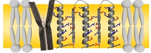
Like the teeth of a zipper, the charged amino acids (red, blue) form connections between protein segments. In this way, they can form pores in the cell membrane. (Figure: KIT)
“It is fascinating to see the elegant basic principles that are used by nature to construct molecular assemblies,” explains Anne Ulrich, Director of the KIT Institute for Biological Interfaces. “A charge zipper between the charged side chains is an entirely unexpected mechanism used by membrane proteins to neutralize their charges such that they can be immersed into hydrophobic cell membranes.”
In the study published now, Ulrich and her team investigate the so-called Twin-arginine translocase (Tat) that is used in the cell membrane of bacteria as an export machinery for folded proteins. Several TatA subunits assemble as a pore that can adapt its diameter to the size of the cargo to be transported. “But how can such a pore be built up from TatA proteins? How can they reversibly form a huge hole in the membrane for a variety of molecules to pass through, but without causing leakage of the cell?”, Ulrich formulates the questions studied.
To answer these questions, the researchers studied the molecular structure of TatA protein from the bacterium B. subtilis, which consists of a chain of 70 amino acids. The analysis showed that it folds into a rather rigid, rod-shaped helix that is followed by a flexible, extended stretch. Many amino acids in the helix and the adjacent stretch carry positive or negative charges. Surprisingly, the sequence of charges on the helix is complementary to those in the adjacent stretch of the protein. When the protein is folded up at the connection point like a pocket knife, positive and negative charges will always meet and attract each other. Hence, the protein links up both of its segments, similar to the interlocking teeth of a zipper.
“The clou is that this binding principle also works with the neighboring proteins,” Ulrich says. Instead of folding up alone, every TatA protein also forms charge zippers with both of its neighbors. Computer simulations showed that this leads to stable and, at the same time, flexible connections between the adjacent molecules. In this way, any number of proteins can be linked together to form an uncharged ring, which thus lines the TatA pore in the hydrophobic membrane. This novel charge zipper principle does not only seem to play a role in protein transport, but also in the attack of certain antimicrobial peptides on bacteria, or in their formation of biofilms as a response to stress.

source :http://www.kit.edu/visit/pi_2013_12526.php

read more

Friday, 18 January 2013

IMP Scientists shed light on the “dark matter” of DNA


In each cell, thousands of regulatory regions control which genes are active at any time. Scientists at the Research Institute of Molecular Pathology (IMP) in Vienna have developed a method that reliably detects these regions and measures their activity. The new technology is published online by Science this week.
Fluorescence image of ovarian tissue of the fruit fly.
 DNA is stained in blue, the activity of enhancers is
represented by the green colour.
Copyright: IMP
Genome sequences store the information about an organism’s development in the DNA’s four-letter alphabet. Genes carry the instruction for proteins, which are the building blocks of our bodies. However, genes make up only a minority of the entire genome sequence – roughly two percent in humans. The remainder was once dismissed as “junk”, mostly because its function remained elusive. “Dark matter” might be more appropriate, but gradually light is being shed on this part of the genome, too.
Far from being useless, the non-coding part of DNA contains so-called regulatory regions or enhancers that determine when and where each gene is expressed. This regulation ensures that each gene is only active in appropriate cell-types and tissues, e.g. haemoglobin in red blood cell precursors, digestive enzymes in the stomach, or ion channels in neurons. If gene regulation fails, cells express the wrong genes and acquire inappropriate functions such as the ability to divide and proliferate, leading to diseases such as cancer.
Despite the importance of gene regulatory regions, scientists have been limited in their ability to study them on a genome-wide scale. Their identification relied on indirect means, which were error prone and required tedious experiments for validating and quantifying enhancer activities..
Alexander Stark and his team at the IMP in Vienna now closed this gap with the development of a new technology called STARR-seq (self-transcribing active regulatory region sequencing), published online by Science this week. STARR-seq allows the direct identification of DNA sequences that function as enhancers and simultaneously measures their activity quantitatively in entire genomes. 
Applying their technology to Drosophila cells, the IMP-scientists surprisingly find that the strongest enhancers reside in both regulatory genes that determine the respective cell-types as well as in broadly active “housekeeping” genes that are required for basic cell survival in most or all cells. In addition, they find several enhancers for each active gene, which might provide redundancy to ensure robustness of gene regulation. 
The new method combines advanced sequencing technology and highly specialized know-how in bio-computing. It is a powerful tool which, according to Alexander Stark, will prove immensely valuable in the future. “STARR-seq is like a magic microscope that lets us zoom in on the regulatory regions of DNA. It will be crucial to study gene regulation and how it is encoded in the genome – both during normal development and when it goes wrong in disease.”
source: http://www.alphagalileo.org/ViewItem.aspx?ItemId=127648&CultureCode=en

read more